Home › Unlabelled › Bone Cross Section Microscope : Cross Section Human Cartilage Bone Under Microscope View Stock Photo Image Of Anatomical Healthcare 128543858 : Both types of bone marrow are enriched with blood vessels and capillaries.2.
Bone Cross Section Microscope : Cross Section Human Cartilage Bone Under Microscope View Stock Photo Image Of Anatomical Healthcare 128543858 : Both types of bone marrow are enriched with blood vessels and capillaries.2.. Jump to navigation jump to search. A neutron can have many types of interactions with a nucleus (ragheb, 2011). Also cross section doesn't have to be circular. This slide showing a cross section of the mammalian trachea (wind pipe) contains examples of several different kinds of tissues. Start studying bone cross section.
Observe that the matrix of the bone is deposited in concentric layers that are called lamellae. Longitudinal & cross sections, 250x shows: We obtained 24 axial slices of the normal brain. Use electromagnets to focus electrons resulting in significantly greater magnifications and resolutions. Learn vocabulary, terms and more with flashcards, games and other study tools.
Mic Uk Microscopy Of Bone from www.microscopy-uk.org.uk Bones protect the various organs of the body, produce red and white blood cells, store minerals. Microscopes of higher quality sometimes use a cross table as stage. Accuracy of the tested digitization method was expressed by. A neutron can have many types of interactions with a nucleus (ragheb, 2011). Learn vocabulary, terms and more with flashcards, games and other study tools. An mri was performed on a healthy subject, with several acquisitions with different weightings: The microscopic cross section represents the effective target area of a single target nucleus for an incident particle. Longitudinal & cross sections, 250x shows:
Cookie information is stored in your browser and performs functions such as recognising you when you return to our website and helping our team to understand which sections of the website you find most interesting and useful.
Picture of cross section human cartilage bone under microscope view for education histology. In a cross section of a bone you can usually see two types of bone tissue what are these called? Accuracy of the tested digitization method was expressed by. Longitudinal & cross sections, 250x shows: The large dark spots are passages for blood vessels and nerves. A polished thin section containing an implant may be used for both transmitted and reflected light examination. The finished bone section will be bonded to a microscope slide and so the first step is to grind flat and polish the part of the bone that will be glued to the slide. They build the entire picture, improve your understanding, consolidate the information and facilitate recall. Also cross section doesn't have to be circular. Bones protect the various organs of the body, produce red and white blood cells, store minerals. A method for preparing samples is described. Under the microscope footage of a transverse section of hard bone. Bone marrow aspiration uses a hollow needle to remove a small sample (about 1 ml) of bone marrow for examination under a microscope.
The implant, which is opaque, is examined by reflected light for features such as porosity, grain size and corrosion. These bone cells have long branching arms (d) which lets them communicate with. Which microscope is used to see the cross section of a stem? Fixed slide cross section of a femur bone cross section human cartilage bone under microscope view for education histology. The large dark spots are passages for blood vessels and nerves.
Rat Rib Transverse Section 250x Spongy Osseous Tissue Osseous Tissue Mammals Skeletal System Other Systems Comparative Anatomy Of Vertebrates Animal Histology Photos from www.nature-microscope-photo-video.com Picture of cross section human cartilage bone under microscope view for education histology. We obtained 24 axial slices of the normal brain. To trace the longer pathways that interconnect different brain regions, cbs labs developed a genetic method to label it is an object of curiosity to know how would human bones look when scanned by an electron microscope. They build the entire picture, improve your understanding, consolidate the information and facilitate recall. When the light that enters the condenser is polarized by placing a polarizer in the filter holder and a second, crossed polarizer at the image plane. Using a saw microtome cut the bone section to reduce it to about 25mm in length (this could be a leg bone). A method for preparing samples is described. From wikimedia commons, the free media repository.
The large dark spots are passages for blood vessels and nerves.
These bone cells have long branching arms (d) which lets them communicate with. Cookie information is stored in your browser and performs functions such as recognising you when you return to our website and helping our team to understand which sections of the website you find most interesting and useful. In a cross section of a bone you can usually see two types of bone tissue what are these called? Longitudinal and cross sections of skeletal muscle fibers (cells), striations, a bands (dark), i bands (light), z lines, myofibrils (longitudinal & cross sectional views), and endomysium. Use electromagnets to focus electrons resulting in significantly greater magnifications and resolutions. The large dark spots are passages for blood vessels and nerves. Stock photo, images and stock. When the light that enters the condenser is polarized by placing a polarizer in the filter holder and a second, crossed polarizer at the image plane. Longitudinal & cross sections, 250x shows: A polished thin section containing an implant may be used for both transmitted and reflected light examination. The microscopic cross section represents the effective target area of a single target nucleus for an incident particle. Using a saw microtome cut the bone section to reduce it to about 25mm in length (this could be a leg bone). Fixed slide cross section of a femur bone cross section human cartilage bone under microscope view for education histology.
Bone marrow aspiration uses a hollow needle to remove a small sample (about 1 ml) of bone marrow for examination under a microscope. A polished thin section containing an implant may be used for both transmitted and reflected light examination. Jump to navigation jump to search. This simply involves placing a section of the bone on the microscope stage and viewing. We obtained 24 axial slices of the normal brain.
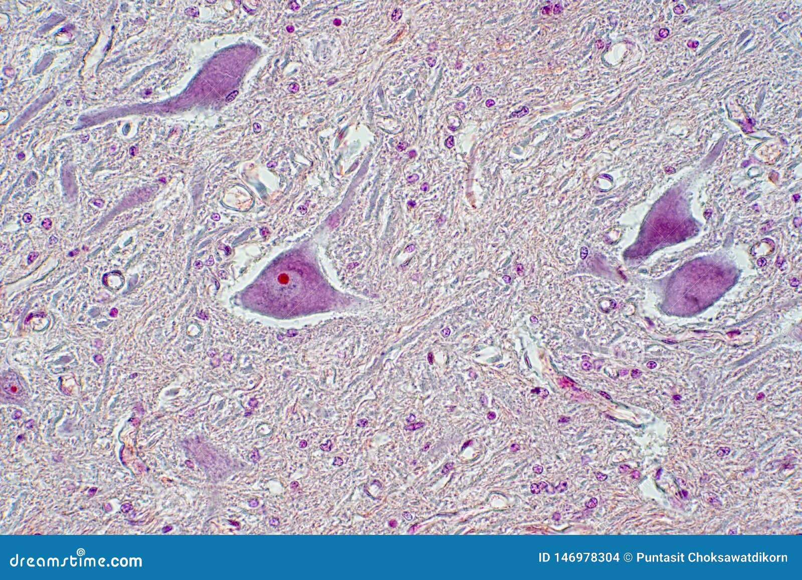
Cross Section Of Spinal Cord Under The Microscope View Stock Photo Image Of Male Bone 146978304 from thumbs.dreamstime.com They build the entire picture, improve your understanding, consolidate the information and facilitate recall. The implant, which is opaque, is examined by reflected light for features such as porosity, grain size and corrosion. Fixed slide cross section of a femur bone cross section human cartilage bone under microscope view for education histology. The finished bone section will be bonded to a microscope slide and so the first step is to grind flat and polish the part of the bone that will be glued to the slide. Accuracy of the tested digitization method was expressed by. Longitudinal & cross sections, 250x shows: Bone marrow aspiration uses a hollow needle to remove a small sample (about 1 ml) of bone marrow for examination under a microscope. Bone cross section — stock image.
Stock photo, images and stock.
The lining of the trachea this model shows a cross section of compact bone. Coloured scanning electron micrograph (sem) of a section through an osteoclast bone cell in reabsorbing bone matrix, showing the cell's nucleus (round, centre). Learn vocabulary, terms and more with flashcards, games and other study tools. When the light that enters the condenser is polarized by placing a polarizer in the filter holder and a second, crossed polarizer at the image plane. This section will examine the gross anatomy of bone first and then move on to its histology. Stock photo, images and stock. Jump to navigation jump to search. Also cross section doesn't have to be circular. We obtained 24 axial slices of the normal brain. From wikimedia commons, the free media repository. Picture of cross section human cartilage bone under microscope view for education histology. Start studying bone cross section. This slide showing a cross section of the mammalian trachea (wind pipe) contains examples of several different kinds of tissues.
Use electromagnets to focus electrons resulting in significantly greater magnifications and resolutions bone cross section. This simply involves placing a section of the bone on the microscope stage and viewing.


Konversi Kode